Interplay between lymphocyte subpopulation, inflammatory cytokines and their correlation with oxidative stress parameters in COVID-19

Submitted: 31 October 2022
Accepted: 13 December 2022
Published: 30 December 2022
Abstract Views: 1010
PDF: 177
HTML: 14
Publisher's note
All claims expressed in this article are solely those of the authors and do not necessarily represent those of their affiliated organizations, or those of the publisher, the editors and the reviewers. Any product that may be evaluated in this article or claim that may be made by its manufacturer is not guaranteed or endorsed by the publisher.

Authors

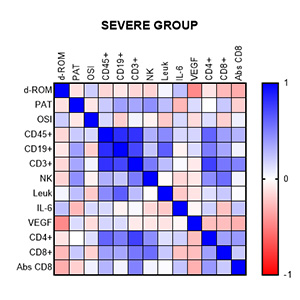
Our objective was to investigate the inflammatory and oxidative stress markers in patients with moderate and severe form of coronavirus disease 2019 (COVID-19). In addition, we show the correlation between changes in lymphocyte subsets and markers of oxidative stress as a tool for patient classification. Interleukin-6 (IL-6) and VEGF were analyzed by utilizing a High Sensitivity Evidence Investigator™ Biochip Array technology. The total antioxidant capacity (PAT) and the free radical concentrations (d-ROM) were measured in serum utilizing analytical photometric system FRAS5. Peripheral blood was used to determine CD45 + mononuclear, B, T, and NK cells using a multi-parameter flow cytometric immunophenotypic test. Statistically significant differences in IL-6 and VEGF levels were observed between the two patient groups. Decreased values of the absolute number of lymphocytes and their CD4 + and CD8 + positive T cells, NK cells, and CD8 were obtained. In the moderate group, good correlations were found between IL-6 and VEGF and NK cells (r=0.6973, P<0.05; for IL-6 and r=0.6498, P<0, for VEGF. 05). Cytokines were correlated with CD45+ (r=0.5610, P<0.05; for IL-6 and r=0.5462, P<0.05 for VEGF). The oxidative stress index can be used as a cheaper alternative and as a triage tool between severe and moderate illnesses, after showing good correlation with more expensive patient classification analysis.

Dimensions

Altmetric

PlumX Metrics

Downloads

Download data is not yet available.

Citations

Huang C, Wang Y, Li X, et al. Clinical features of patients infected with 2019 novel coronavirus in Wuhan, China. Lancet 2020;395:497-506.
Zhou P, Yang XL, Wang XG, et al. A pneumonia outbreak associated with a new coronavirus of probable bat origin. Nature 2020; 579:270-3.
Lange A, Lange J, Jaskuła E. Cytokine overproduction and immune system dysregulation in allo HSCT and COVID-19 patients. Front Immunol 2021;12:658896.
Xu Z, Shi L, Wang Y, et al. Pathological findings of COVID-19 associated with acute respiratory distress syndrome. Lancet Respir Med 2020;8:420-2.
Ramljak D, Vukoja M, Curlin M, et al. Early Response of CD8+ T Cells in COVID-19 Patients. J Pers Med 2021;11:1291.
De Zuani, Laznickova P, Tomaskova V, et al. High CD4-to-CD8 ratio identifies an at-risk population susceptible to lethal COVID-19. Scand J Immunol 2022;95:e1312.
Pallotto C, Suardi LR, Esperti S, et al. Increased CD4/CD8 ratio as a risk factor for critical illness in coronavirus disease 2019 (COVID-19): a retrospective multicentre study. Infect Dis (Lond) 2020;52:675-7.
Zhang H, Wu T. CD4+T, CD8+T counts and severe COVID-19: a meta-analysis. J Infect 2020;81:e82-4.
Mahmoudi S, Yaghmaei B, Sharifzadeh Ekbatani M, et al. Effects of coronavirus disease 2019 (COVID-19) on peripheral blood lymphocytes and their subsets in children: imbalanced CD4(+)/CD8(+) T cell ratio and disease severity. Front Pediatr 2021;9:643299.
Rezaei M, Marjani M, Mahmoudi S, et al. Dynamic changes of lymphocyte subsets in the course of COVID-19. Int Arch Allergy Immunol 2021;182:254-62.
Ganji A, Farahani I, Khansarinejad B, et al. Increased expression of CD8 marker on T-cells in COVID-19 patients. Blood Cells Mol Dis 2020;83:102437.
Liu J, Li S, Liu J, et al. Longitudinal characteristics of lymphocyte responses and cytokine profiles in the peripheral blood of SARS-CoV-2 infected patients. EBioMed 2020;55:102763.
Moratto D, Chicarini M, Giustini V, et al. Flow cytometry identifies risk factors and dynamic changes in patients with COVID-19. J Clin Immunol 2020;40:970-3.
Iannetta M, Buccisano F, Fraboni D, et al. Baseline T-lymphocyte subset absolute counts can predict both outcome and severity in SARS-CoV-2 infected patients: a single center study. Sci Rep 2021;11:12762.
Delgado-Roche L, Mesta F. Oxidative stress as key player in severe acute respiratory syndrome coronavirus (SARS-CoV) infection. Arch Med Res 2020;51:384-7.
Petrushevska M, Zendelovska D, Atanasovska E, et al. Presentation of cytokine profile in relation to oxidative stress parameters in patients with severe COVID-19: a case-control pilot study. F1000Res 2021;10:719.
Cecchini R, Cecchini AL. SARS-CoV-2 infection pathogenesis is related to oxidative stress as a response to aggression. Med Hypotheses 2020;143:110102.
World Medical Association. World Medical Association Declaration of Helsinki: ethical principles for medical research involving human subjects. JAMA 2013;310:2191-4.
Panovska-Stavridis I, Trajkova S, Ivanovski M, et al. Minimal screening analysis-based algorithm for diagnosis and clinical stratification of patients with acute myeloid leukaemia (AML): single center experience. Prilozi 2012;33:93-106.
Akbari H, Tabrizi R, Lankarani KB, et al. The role of cytokine profile and lymphocyte subsets in the severity of coronavirus disease 2019 (COVID-19): A systematic review and meta-analysis. Life Sci 2020;258:118167.
Zendelovska D, Atanasovska E, Petrushevska M, et al. Evaluation of oxidative stress markers in hospitalized patients with moderate and severe COVID-19. Rom J Intern Med 2021;59:375-83.
Han H, Ma Q, Li C, et al. Profiling serum cytokines in COVID-19 patients reveals IL-6 and IL-10 are disease severity predictors. Emerg Microbes Infect 2020;9:1123-30.
Alkharsah KR. VEGF upregulation in viral infections and its possible therapeutic implications. Int J Mol Sci 2018;19:1642.
Huet T, Beaussier H, Voisin O, et al. Anakinra for severe forms of COVID-19: a cohort study. Lancet Rheumatol 2020;2:e393-e400.
Lu C, Liu Y, Chen B, et al. Prognostic value of lymphocyte count in severe COVID-19 patients with corticosteroid treatment. Signal Transduct Target Ther 2021;6:106.
Bogdanic N, Lukic Lj, Begovac J. Coronavirus disease 19 (COVID-19) pharmacologic treatment: Where are we now? Infektol Glasn 2020;40:134-47.
Malgie J, Schoones JW, Pijls BG. Decreased mortality in coronavirus disease 2019 patients treated with tocilizumab: A rapid systematic review and meta-analysis of observational studies. Clin Infect Dis 2020;ciaa1445.
Balzanelli MG, Distratis P, Dipalma G, et al. Immunity profiling of COVID-19 infection, dynamic variations of lymphocyte subsets, a comparative analysis on four different groups. Microorganisms 2021;9:2036.
de Las Heras N, Martín Giménez VM, Ferder L, et al. Implications of oxidative stress and potential role of mitochondrial dysfunction in COVID-19: therapeutic effects of vitamin D. Antioxidants (Basel) 2020;9:897.
Alamdari DH, Moghaddam AB, Shahram Amini S, et al. Application of methylene blue -vitamin C - N-acetyl cysteine for treatment of critically ill COVID-19 patients, report of a phase-I clinical trial. Eur J Pharmacol 2020;885:173494.

How to Cite

Petrusevska, M., Zendelovska, D., Atanasovska, E., Spasovska, K., Grozdanovski, K., Stojanovska, S., Panovska-Stavridis, I., & Eftimov, A. (2022). Interplay between lymphocyte subpopulation, inflammatory cytokines and their correlation with oxidative stress parameters in COVID-19. Italian Journal of Medicine, 16(1). https://doi.org/10.4081/itjm.2022.1543